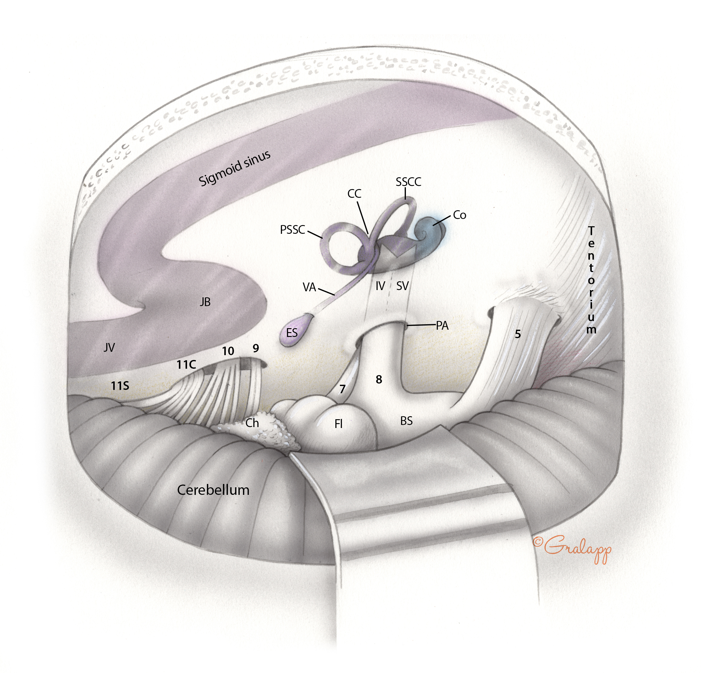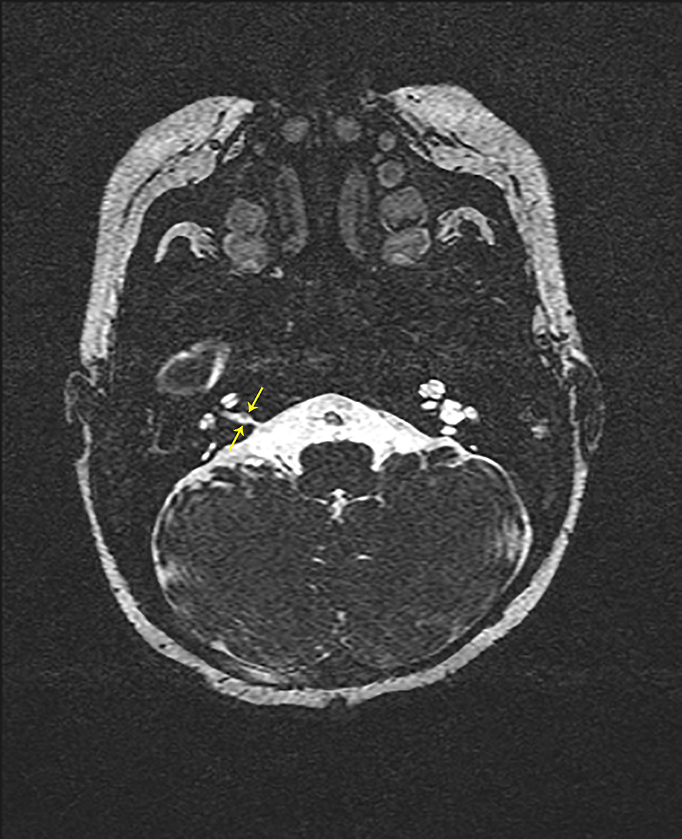
IAC enhancement was ipsilateral to the facial hemangioma in 7/7 patients.

Follow-up studies were available on 4 of these 7 patients, all of which demonstrated resolution of enhancement or reduction in lesion size ( Fig 5). Asymmetric IAC enhancement without a discrete mass was identified in 1 patient. Of the 15 patients in whom MR imaging was performed with contrast during the first 3 years of life, 6 had avidly enhancing IAC masses consistent with hemangiomas. One neonate did not receive intravenous contrast. Of the remaining 4 patients, 3 were initially imaged with intravenous contrast at ages 8, 17, and 18 years. One patient with enlargement of the left IAC had bilateral facial hemangiomas.įifteen of 19 patients with unilateral IAC enlargement underwent contrast-enhanced thin-section imaging through the posterior fossa during the first 3 years of life. IAC enlargement was ipsilateral to the facial hemangioma in 17/19 patients and contralateral in 1/19. Nineteen of 44 (43%) cases demonstrated unilateral funnel-shaped IAC enlargement this group of 19 patients included 16 female and 3 male patients between 8 days and 18 years of age (average age, 40 months) at the time of initial MR imaging. All abnormal findings were classified as ipsilateral or contralateral to the patients' facial hemangiomas ( Fig 4). All MR imaging studies were evaluated for asymmetric caliber or contour of the IAC, intracanalicular enhancement, cerebellar asymmetry, and asymmetric size or morphology of the petrous and occipital bones ( Fig 3). Multiplanar postgadolinium T1-weighted images were acquired at a 3- to 5-mm section thickness with a 0- or 1-mm gap, with or without a fat-saturation technique. Typically, axial T2-weighted images were obtained by using an FSE or TSE technique at section thicknesses ranging from 2.5 to 5 mm, with a 0- or 1-mm gap. MR imaging techniques varied slightly among the patients, depending on the location of their scans and whether the brain examinations were modified for inclusion of imaging of the face to evaluate facial hemangiomas. A neuroradiologist with 9 years of experience and a pediatric neuroradiologist with 20 years of experience performed a consensus review of MR imaging examinations of the brain from pediatric patients with an established diagnosis of PHACES association based on accepted clinical criteria.

Institutional review board approval was obtained from the 2 participating institutions for this retrospective review. Subsequent review of clinical data bases at 2 institutions identified 44 patients (37 male, 7 female) diagnosed with PHACES association who underwent diagnostic MR imaging of the brain between 20. We raise the possibility of an association between enlargement of the internal auditory canal in PHACES and a generalized malformation of the posterior fossa with cerebellar and calvarial hypoplasia.

Imaging was reviewed for abnormal enhancement in the internal auditory canal, internal auditory canal enlargement, cerebellar hypoplasia, prominence of the petrous ridge, and deformity of the calvarium. We reviewed our records to identify children with PHACES association who had been evaluated with MR imaging at our institutions. SUMMARY: We noted enlargement of the internal auditory canal in several of our patients with posterior fossa malformations, hemangiomas, arterial anomalies, cardiac defects, eye abnormalities, and sternal or supraumbilical defects (PHACES) association and hence evaluated children with PHACES for the presence of an enlarged internal auditory canal and potential associated findings, including infantile hemangioma within the internal auditory canal, to understand the genesis of this enlargement.


 0 kommentar(er)
